For the analysis of cynomolgus monkey (Macaca Fascialis) brain data the Cynomolgus_CIMA-UN VOI atlas [1] is available. We would like to thank Elena Prieto and Maria Collantes from the Centro de Investigacion Medica Aplicada (CIMA), Universidad de Navarra, for providing the data and helping with the integrations.
Spatial Normalization
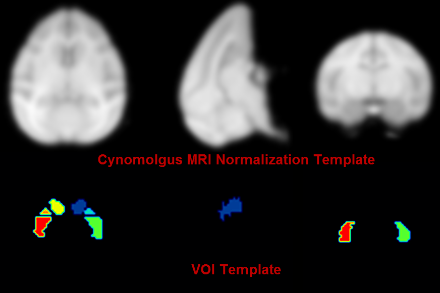
A T1-weighted MRI normalization template created from 15 healthy animals is available as Cynomolgus_CIMA-UN-MRI. Two PET templates in the same space are also provided: Cynomolgus_CIMA-UN-Dopa and Cynomolgus_CIMA-UN-DTBZ.
VOI Atlas
The VOIs were hand drawn in the striatum (VOI size 400 mm3) and occipital lobe (VOI size 310 mm3) on axial MRI slices based on anatomical borders.
Reference
1.Collantes M, Prieto E, Penuelas I, Blesa J, Juri C, Marti-Climent JM, Quincoces G, Arbizu J, Riverol M, Zubieta JL, Rodriguez-Oroz MC, Luquin MR, Richter JA, Obeso JA. New MRI, 18F-DOPA and 11C-(+)-alpha-dihydrotetrabenazine templates for Macaca fascicularis neuroimaging: advantages to improve PET quantification. Neuroimage. 2009;47(2):533-9. DOI