“Centiloid” was introduced as a “standard” method for the quantification of the amyloid load by Klunk et al. [1]. It uses a lumped SUVr value obtained with PiB PET as the standard, and describes a scaling method to transform the outcome using other amyloid PET tracers into a comparable measure.
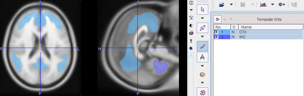
The lumped SUVr is defined by a cortical target region CTX and a reference region in the MNI space. CTX is a data-driven cortex VOI which includes the typical brain regions with high amyloid load in Alzheimer's Disease including the frontal, temporal and parietal cortices, precuneus, the anterior striatum and insular cortex. While different reference regions were tested, use of whole cerebellum WC is the final recommendation.
The Centiloid atlas in PMOD only includes the CTX and WC VOIs, as illustrated below on top of the ICBM152T1 template. Note that the locations are correct, although they don't follow the anatomical boundaries. This behavior is due to the PET data-driven process for their derivation, and has been confirmed by the authors.
Reference
1.Klunk WE, Koeppe RA, Price JC, Benzinger TL, Devous MD Sr, Jagust WJ, Johnson KA, Mathis CA, Minhas D, Pontecorvo MJ, Rowe CC, Skovronsky DM, Mintun MA. The Centiloid Project: standardizing quantitative amyloid plaque estimation by PET. Alzheimers Dement. 2015; 11(1):1-15.e1-4. DOI: