Overview
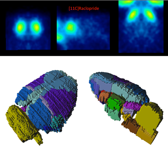
The Px Rat (A.Schwarz) atlas is based on 97 anatomical MR images of adult male Sprague-Dawley rats (250-300g). For the original atlas [1] a volumetric reconstruction of the Paxinos and Watson rat brain atlas was created and adapted to the averaged anatomy. This version of the atlas was used as part of a proposed standardized methodology for the creation of small animal brain PET templates [2]. For application in PMOD the atlas and templates were interpolated to 0.1 mm resolution and VOIs merged to avoid small regions, which would result in poor statistics. The VOI atlas contains 60 cortical and subcortical regions. The atlas is distributed with PMOD by courtesy of Dr. Adam Schwarz, Department of Psychological and Brain Sciences, Indiana University, and the PET/SPECT templates courtesy of University Medical Center Groningen, The Netherlands (with thanks to Dr. D. Vallez Garcia, UMC Groningen, Nuclear Medicine & Imaging).
Spatial Normalization
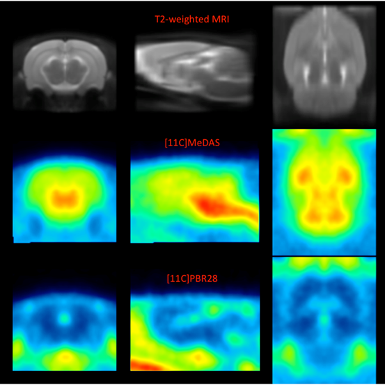
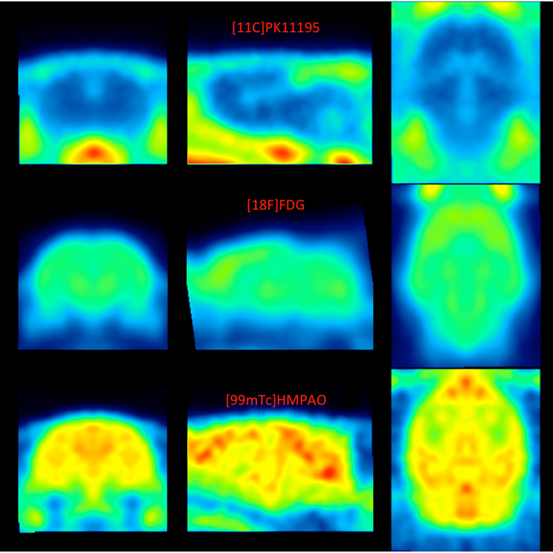
The T2-weighted MR template, five PET templates and a single SPECT template are available in the Fuse It tool when Rat is selected as Species.
▪Px Rat (A.Schwarz)-T2, Px Rat (Groningen)-T2: This is the T2-weighted MR anatomical reference for the A.Schwarz VOI atlas.
▪Px Rat (Groningen)-MeDAS: This is a PET template for the tracer [11C]MeDAS, coregistered to the MR anatomical reference above.
▪Px Rat (Groningen)-PBR28: This is a PET template for the tracer [11C]PBR28, coregistered to the MR anatomical reference above.
▪Px Rat (Groningen)-PK11195: This is a PET template for the tracer [11C]PK11195, coregistered to the MR anatomical reference above.
▪Px Rat (Groningen)-Raclopride: This is a PET template for the tracer [11C]Raclopride, coregistered to the MR anatomical reference above.
▪Px Rat (Groningen)-FDG: This is a PET template for the tracer [18F]FDG, coregistered to the MR anatomical reference above.
▪Px Rat (Groningen)-SPECT: This is a SPECT template for the tracer [99mTc]HMPAO, coregistered to the MR anatomical reference above.
The image files corresponding to these templates can be found in the resources/templates/voitemplates/Px Rat (A.Schwarz) and resources/templates/voitemplates/Px Rat (Groningen) folders, specifically in the normalization sub-folder. Mask files for use during normalization and coregistration are also available.


VOI Atlas
The VOI atlas Px Rat (A.Schwarz) (and identical atlas through Px Rat (Groningen) ) can be selected in the list of included VOI atlases. The corresponding map files in Nifti format can be found in the resources/templates/voitemplates/Px Rat (A.Schwarz) directory.
The brain VOIs are structurally organized in a tree on the Group tab of the VOI editing page. The selection of a VOI subset is supported by a dedicated user interface.
Reference
1.Schwarz AJ, Danckaert A, Reese T, Gozzi A, Paxinos G, Watson C, Merlo-Pich EV, Bifone A. A stereotaxic MRI template set for the rat brain with tissue class distribution maps and co-registered anatomical atlas: application to pharmacological MRI. Neuroimage. 2006 Aug 15;32(2):538-50. DOI.
2.Vallez Garcia D, Casteels C, Schwarz AJ, Dierckx RA, Koole M, Doorduin J. A standardized method for the construction of tracer specific PET and SPECT rat brain templates: validation and implementation of a toolbox. PLoS One. 2015;10(3):e0122363. DOI.