The result of matching is shown on the MATCHED PET page. Please verify that matching was successful by evaluating the alignment in different parts of the brain. Particularly helpful to do so is to interactively drag the fusion balance left/right, and to enable contour outlines.
If the match is not satisfactory, there are two options to rectify the situation:
1.Return to the previous page, change the sampling and smoothing parameters and try the automatic matching again, or
2.Activate the Adjust matching button and shift/rotate the PET image interactively by dragging the handles in the image or entering offsets/angles on the Move/Rotate tabs. Finally the transformation needs to by applied with the ![]() button.
button.
Normalization
The next step is to spatially normalize the subject images. There are three options as illustrated below.
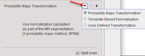
Probability Maps Transformation |
Uses the normalization resulting from the GM/WM/CSF MR segmentation procedure. |
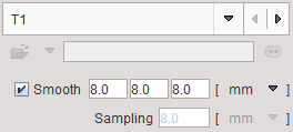
Template Based Normalization |
Performs an SPM5-type normalization between the subject MR and the T1 atlas template image with the usual options |
User Defined Transformation |
For loading and applying a normalization transformation which has previously been calculated for the MR image and saved. |
The Split brain option is only relevant for the separation of the white matter into left hemispheric, right hemispheric, and cerebellar parts. If the procedure is not enabled or fails, then a global white matter region is created.
Note: The brain split division uses a template in the MNI space. This template consists of three labels corresponding to the left hemisphere, the right hemisphere and the cerebellar part. In the brain split process the brain mask is eroded with an initial erosion size and the separation into individual segments is verified. The result must consist in at least 3 separate segments. If it is not the case the erosion size is incremented with the erosion step. The algorithm verifies each separate region to which label in the template belongs. The front propagation is used to divide the whole brain mask only when all the three region seeds are found to be geodesic.
Please activate the Normalize action button to proceed.