The stereotactic normalization result is shown on the NORMALIZED page. Please verify that the normalization procedure was successful by evaluating the alignment in different parts of the brain. Particularly helpful are the iso-contour lines. The thresholds on the TEMPLATE and the NORMALIZED should be adjusted so that the lines follow the same structures. The final brain structure outlines will only be adequate if the normalization succeeded.
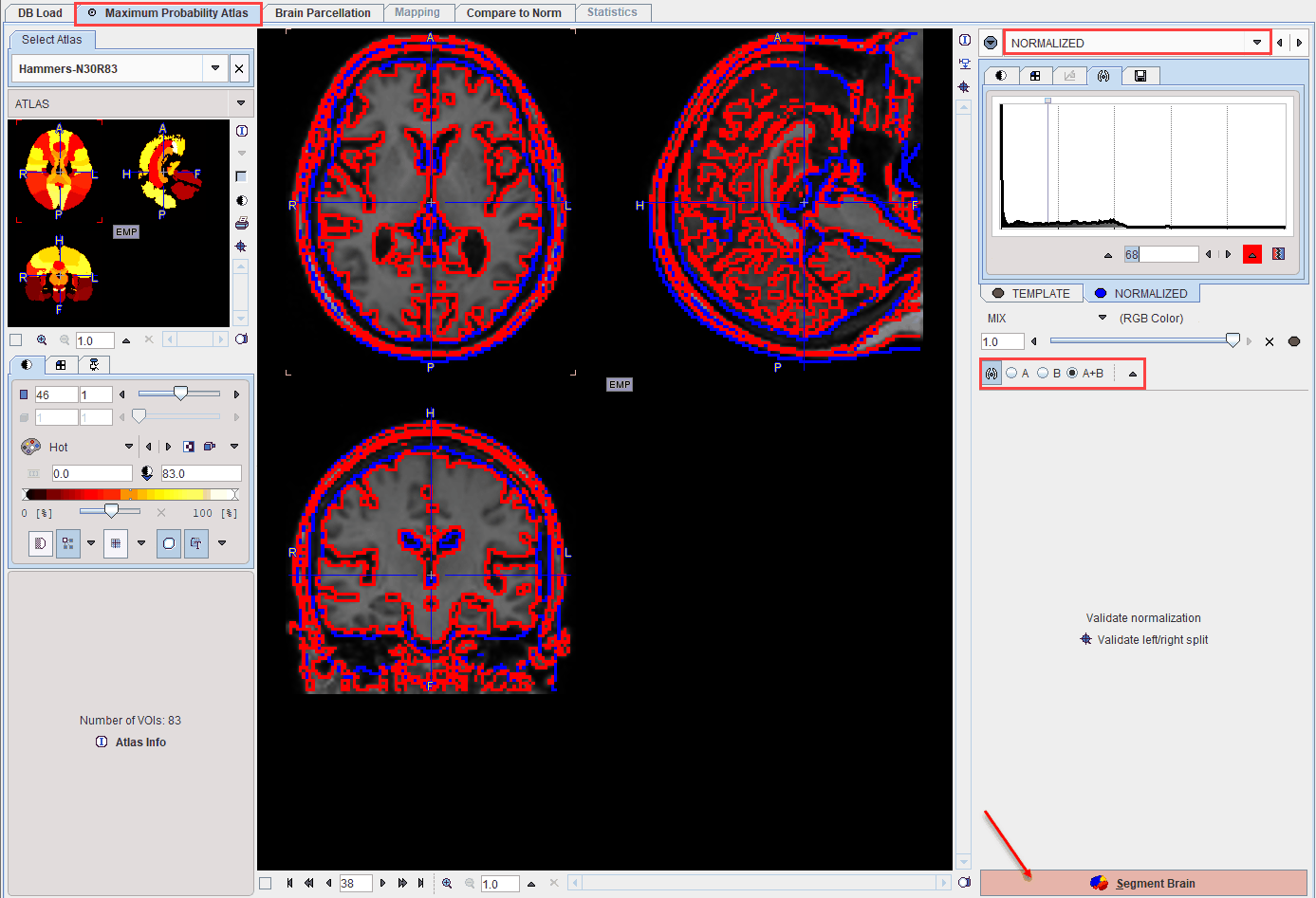
After successful normalization the mapping between the different image spaces is established:
▪the normalization maps the MR to the atlas space;
▪the rigid transform maps the PET to MRI space;
▪the rigid transform combined with the normalization results in a transform which maps the PET to the atlas space.
As all the transformations can be inverted, the atlas space can also be mapped to PET and the MR image space. Consequently, the brain structures which are defined in the atlas space can be mapped to the PET and MR subject space and shown in the overlay.
Left/Right Split Validation
The Validateleft/right split button opens a dialog window for inspecting the result of the brain splitting procedure.
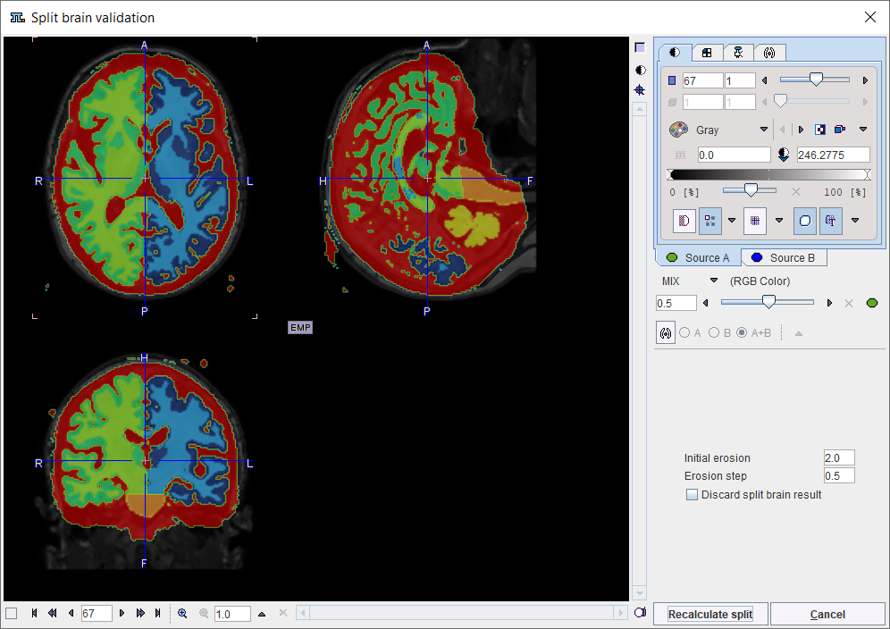
There are 7 resulting segments, with are shown in the Source B tab. They correspond to left white matter, left gray matter , right white matter, right gray matter, cerebellar gray matter (including pons), cerebellar white matter and CSF. If the procedure doesn't come up with a reasonable splitting into right and left parts, there are two options to proceed. The Initial erosion size and Erosion step parameters can be modified and the procedure restarted using Recalculate split, or splitting can be switched off by the Discard split brain result flag. In the latter case please confirm with the Discard split button to close the window.
Atlas Selection
The Maximum Probability Atlas module is aimed at leveraging the brain structures defined in the N30R83 atlas. However, the same workflow can also be applied to other atlases, even to atlases defined for other species. For the selection of the atlas please activate the blue show/hide button left to NORMALIZED.
![]()
As a consequence, a window appears to the left of the page which allows selecting the atlas from a list. To hide the atlas selection window please activate the show/hide button again.
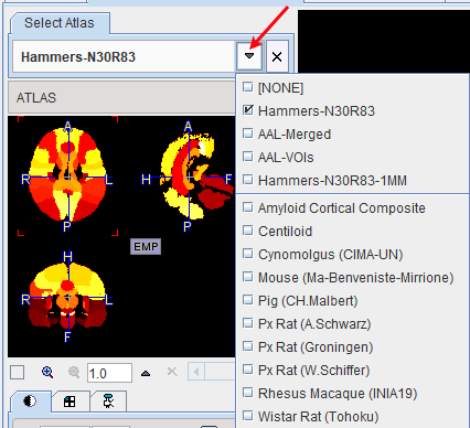
For each of the selections, the ATLAS (defining the structures), the TEMPLATE (pattern for the spatial normalization) and the MASK of the atlas can be inspected.
Please activate the Segment Brain action button to start the mapping of the brain structures of the selected atlas.