Standard Uptake Values (SUV)
The information contained in PET and SPECT images is related to the physical concentration of tracer in tissue. If all images distortions are corrected by the reconstruction procedure, the value units are activity concentrations, for instance Bq/ml. The uptake in tissue is dependent on many factors, but it is directly proportional to the injected activity, and inversely related to the mass within the tracer can distribute. To calculate a measure of tracer uptake which is better comparable among subjects ("Standard Uptake Value" SUV) the measured activities are therefore divided by the injected dose and multiplied by the body mass.
SUV Calculation Methods
Different variants of the SUV calculation are used. The most straightforward directly uses the subject weight entered in the demographic subject information. Because tracer uptake in fat is typically low, normalization to lean body mass has been found to be a preferable measure. However, recently it has been found that the lean body mass calculation using the James method breaks down for obese subjects, and the Janmahasatian method should be used instead [1]. An additional variant is normalization to the body surface area. A variant used in preclinical imaging is normalization to the injected dose.
The formula applied by the PMOD SUV calculation are:
1.SUV Body weight [g/ml] = (A/D)*W*1000.
2.SUV Lean Body Mass [g/ml] James method (normal BMI range) = (A/D)*LBM*1000.
LBM(Female) = 1.07*W-148*(W/H)2,
LBM(Male) = 1.10*W-128*(W/H)2.
3.SUV Lean Body Mass [g/ml] Janmahasatian method (also for extreme BMI) = (A/D)*LBM*1000.
LBM(Female) = 9270*W/(8780 + 244*W/L2).
LBM(Male) = 9270*W/(6680 + 216*W/L2).
4.SUV Body Surface Area [cm2/ml] = (A/D)*BSA*10000.
BSA = (W0.425 x H0.725) x 0.007184.
5.Injected dose per ml [%ID/ml] = A/D*100.
The variables in the formula are defined as follows:
A |
Activity concentration in the image [Bq/ml]. Note that it has to be decay corrected during reconstruction to a common time for all acquisitions of a whole-body or dynamic scan. |
D |
Applied dose [Bq] at the time the image is corrected to, see below. |
W |
subject weigth [kg] |
H |
subject height [cm] |
L |
subject height [m] |
Usually, the activity of the dose to be applied is measured at a time before injection. Therefore, PMOD needs the following information for calculating the effectively injected dose at the time of image decay correction:
•the half-life of the radionuclide;
•the time when the activity was calibrated before injection;
•the activity remaining in the syringe after injection and when it was calibrated (only available in GE data);
•the time of image decay correction.
The activity remaining in the syringe will be subtracted from the calibrated activity, taking the different decay times into account.
PMOD tries to extract the information needed from the image header. For various reasons (information entered by technician, file format, manufacturer) however, it may be incomplete. In this case the information has to be entered or corrected manually before the SUV can be calculated.
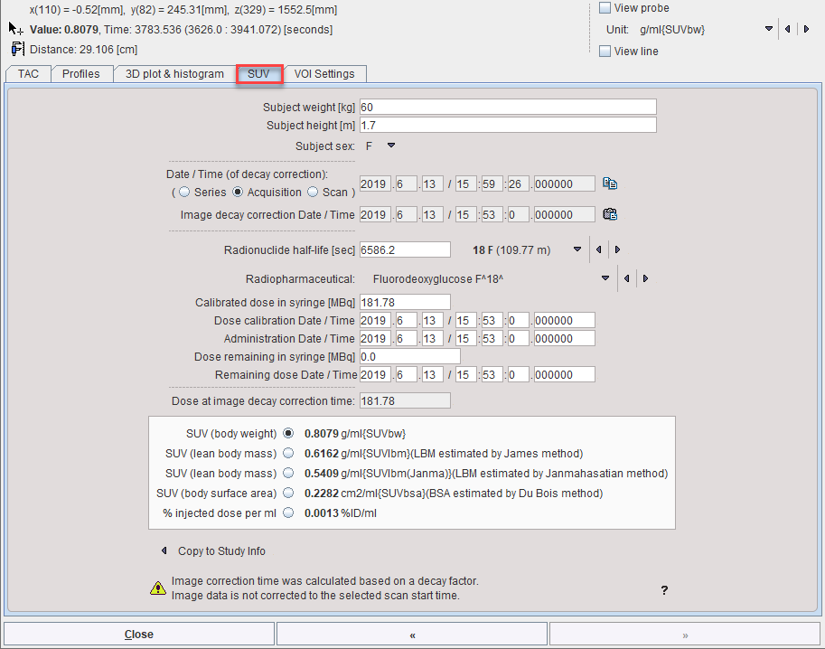
SUV Value Inspection Window
The SUV (Standard Uptake Value) button
![]()
below the data inspector button is a shortcut for opening the data inspector with the SUV tab selected

The window can be extended to show the SUV-related elements with the >> button.

The upper part of the SUV panel shows the elements which are relevant for the SUV calculation. They should be essentially be self-explanatory. The lower part shows the different SUVs of the current pixel. The SUV values change whenever the cursor is moved over the image.
SUV Parameter Details
Unfortunately, the information regarding the time point of decay correction is unreliable in the images. In cases when it is explicitly encoded, it is shown in the Image decay correction Date / Time field. Otherwise, the Date / Time option should be specified such that the time shown represents the image decay correction time. Per default, it is set to the Acquisition time field of DICOM, which however is not always correct. Therefore, it is possible to alternatively select the Series or the Scan time DICOM information. As a convenience the Copy to all button copies the selected Date / Time to all other date and time fields and no correction will be applied to the specified dose.
Series Date/Time |
Abstract time point which defines zero for the time vector of a dynamic series. |
Acquisition Date/Time |
The actual time the data acquisition started (default setting). |
Scan time |
Private GE field; should be used for SUV calculations with other GE fields. |
Image decay correction Date/Time |
May be contained in Enhanced PET objects, a copy of Administration time, calculated from decay factors in some situations, or copied from the selected Date / Time. |
Administration Date/Time |
Standard DICOM time at which radiopharmaceutical administration started. According to the DICOM standard PET images can be also decay corrected to this time point. (Please note that in this case the SUV values will change compared to previous version as an additional factor for the delay from the administration to the scan start will be calculated.) |
Calibrated dose |
Private GE fields for SUV calculations. They are set to the standard total dose and administration time for non GE data. |
Dose remaining... |
Private GE fields for SUV calculations. Are set to 0.0 and Administration time for non GE data. |
Note: Because of the uncertainty of the SUV-related information in DICOM it is recommended to verify PMOD's SUV values with the values calculated by software of the hardware vendor.
Image Display in SUV Units
If all the information required for SUV calculation is present, the display units can be switched from the original activity concentration units to SUV values using the button near the lower color table threshold.
SUV Statistics
The calculation of statistics in SUV units is directly supported on the original image by the VOI functionality, provided that all required information is available. Explicit SUV images can be calculated with the SUV Image Calculation external tool.
Reference
1.Tahari AK, Chien D, Azadi JR, Wahl RL: Optimum lean body formulation for correction of standardized uptake value in PET imaging. J Nucl Med 2014, 55(9):1481-1484. PMC