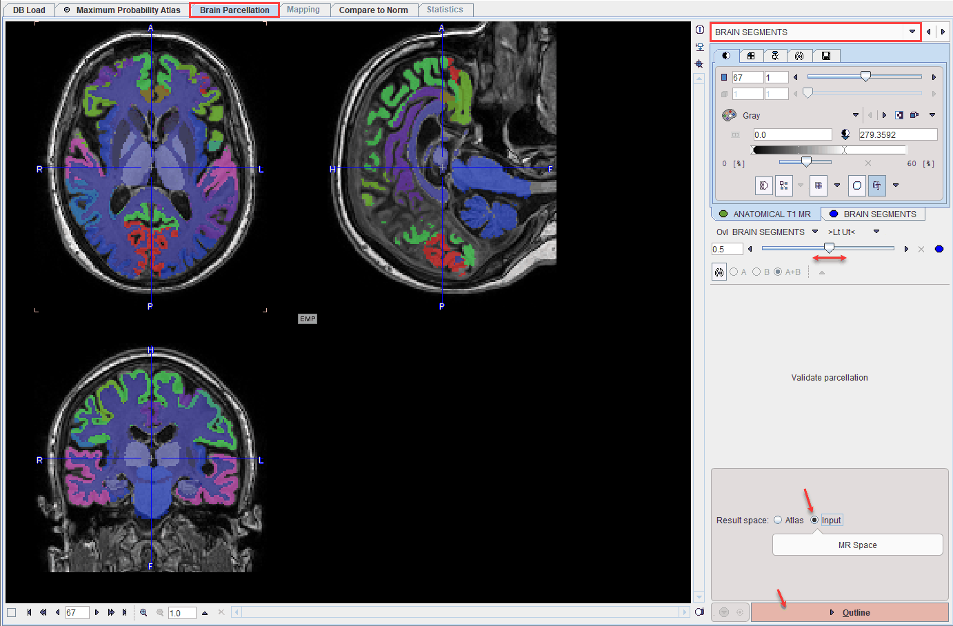
The BRAIN SEGMENTS page shows the parcellation result as an overlay to the MR image.

The BRAIN SEGMENTS image represents a label atlas of the brain structures identified by the segmentation algorithm. Please check the alignment of the structure information with the subject image with the fusion slider. In the case of a mismatch, try changing the GM segment definition on the (previous) TISSUE SEGMENTS page and/or the number of subjects included, and repeat parcellation. Alternatively, the structure definitions can be adjusted manually after the outlining step.
The next step consists of outlining the brain segments in the desired Result space :
Atlas |
The MR image is transformed into the Atlas space and statistics are calculated with interpolated MR values. The Preserve total amount option corrects for volume changes during the spatial normalization step. Image intensities are scaled by the amount of contraction that has occurred during spatial normalization, so that the total amount of gray matter remains the same as in the original image. This option should be used when the normalized MR image is used for voxel-based morphometry (VBM). |
Input |
The statistics are calculated with original MR values. |
Outlining of the brain structures is started with the Outline button.