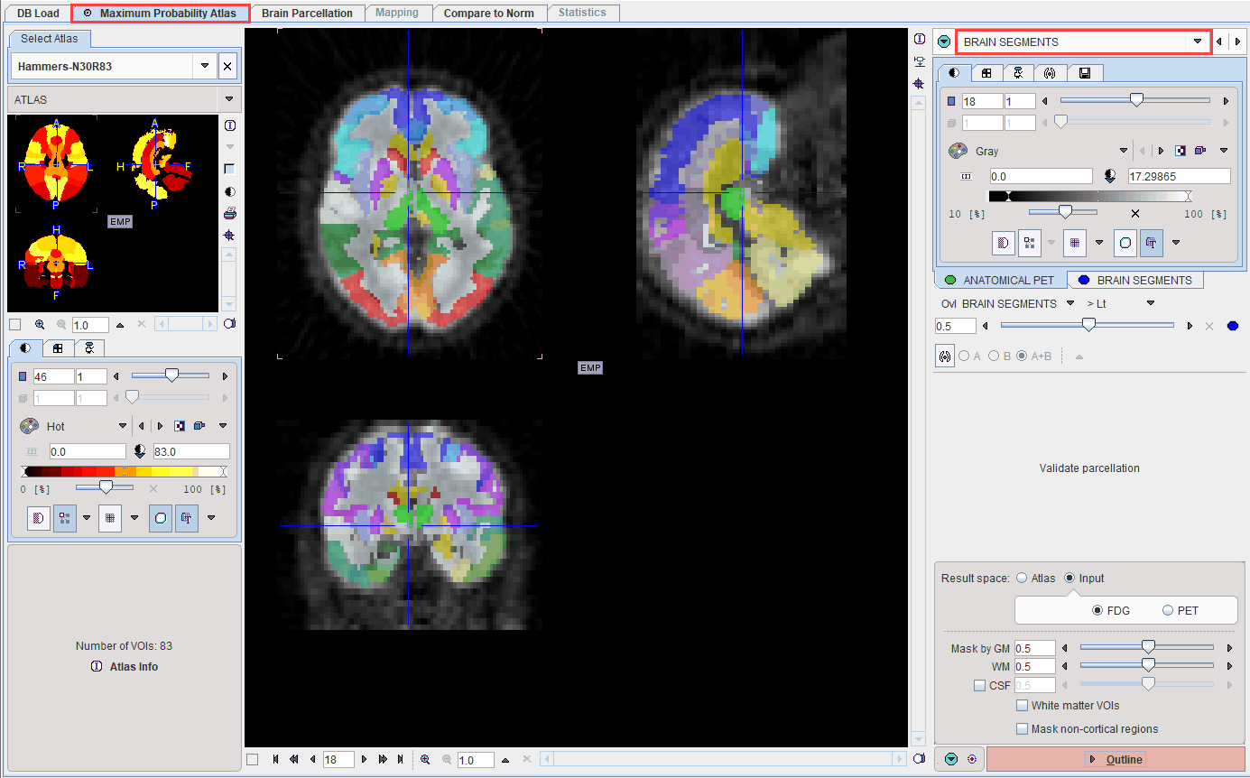
The result of the brain structure transformation is shown on the BRAIN SEGMENTS page. It shows the transformed atlas together with the anatomical PET.
Result Space
There are three options for evaluating the PET image. These can be configured using the Result space radio buttons. The information shown on the page is updated as soon as the configuration is changed. It shows the PET image in the selected result space together with the brain structures.
The Result space options are:
1.Atlas: With this setting the PET image is transformed to the MNI space and the original structures of the N30R83 atlas can be applied to it.
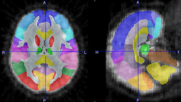
2.Input, FDG: With this setting the atlas structures are transformed to the anatomical PET space.
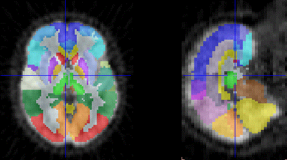
3.Input, PET: With this setting the atlas structures are transformed to the (original) functional PET space.
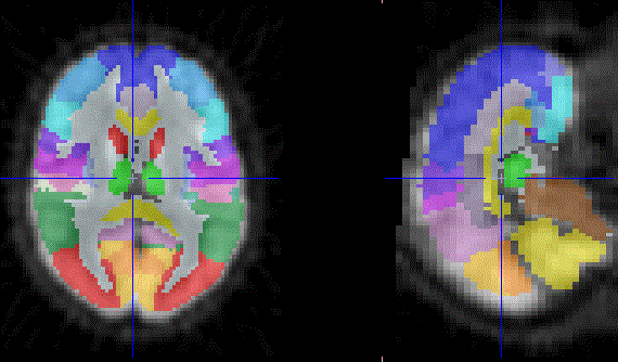
Note that due to the relatively large size of typical PET voxels, the structure outlines are likely to appear pixelated. To avoid this effect the PET image should be reconstructed with small pixel sizes.
Intersection of Atlas Structures with Gray Matter
For atlases with population tissue probability maps users can take advantage of restricting the structures to pixels with a high probability of belonging to gray matter.
![]()
The Mask by GM slider allows the lower threshold to be applied to the GM probability map for creating the mask to be defined. The higher the probability threshold, the thinner the cortical structures become. The illustration below shows masking in the PET space at three increasing GM probability thresholds (CSF box disabled and WM slider set to 1).
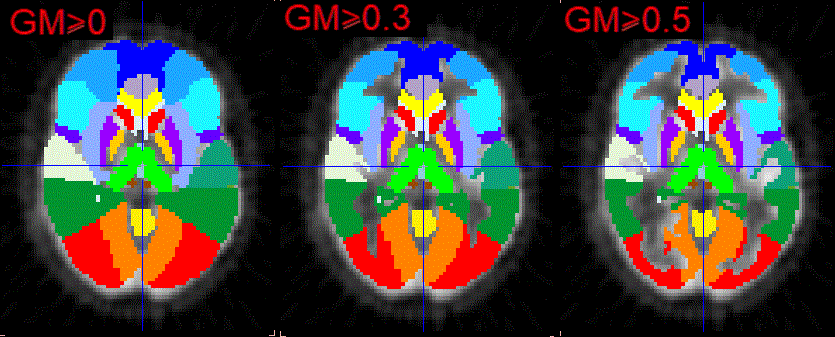
Note that the central structures were not affected by masking in the example above, because the box Mask non-cortical regions was not enabled. If this option is enabled, the central structures also shrink. However, because of the low probability levels in that area, the reduction may become too severe. Therefore, masking of non-cortical regions is disabled by default.
The white matter tissue probability map also can be thresholded at a certain theshold in order to generate a white matter VOI (or VOIs, if White matter segmentation was activated).
With the CSF box enabled, the CSF threshold slider allows further trimming of the GM mask: the higher the threshold the broader the cortical structures become.The illustration below shows masking in the PET space at three decreasing CSF probability thresholds while the GM probability threshold is set to 0.5 and WM probability is set to the value of 1.
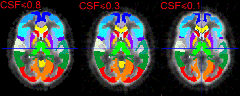
For the atlases with population tissue probability maps the white matter tissue probability map can be used to allow the intersection of the population white matter region with the selected atlas. This can be easily achieved enabling the White matter VOI box.
Structure Outlining
Once the result space and gray matter masking have been specified, the brain structures are fully defined and can be outlined to create contour volumes-of-interest. This process is started with the Outline action button.